Pedigree Chart with Genotypes: Rules, Phenocopies, and Software Guide
Master pedigree charts with genotypes. Learn the rules for autosomal and sex-linked alleles, how to handle phenocopies and negative tests, and generate genotype pedigrees in Biogistry.
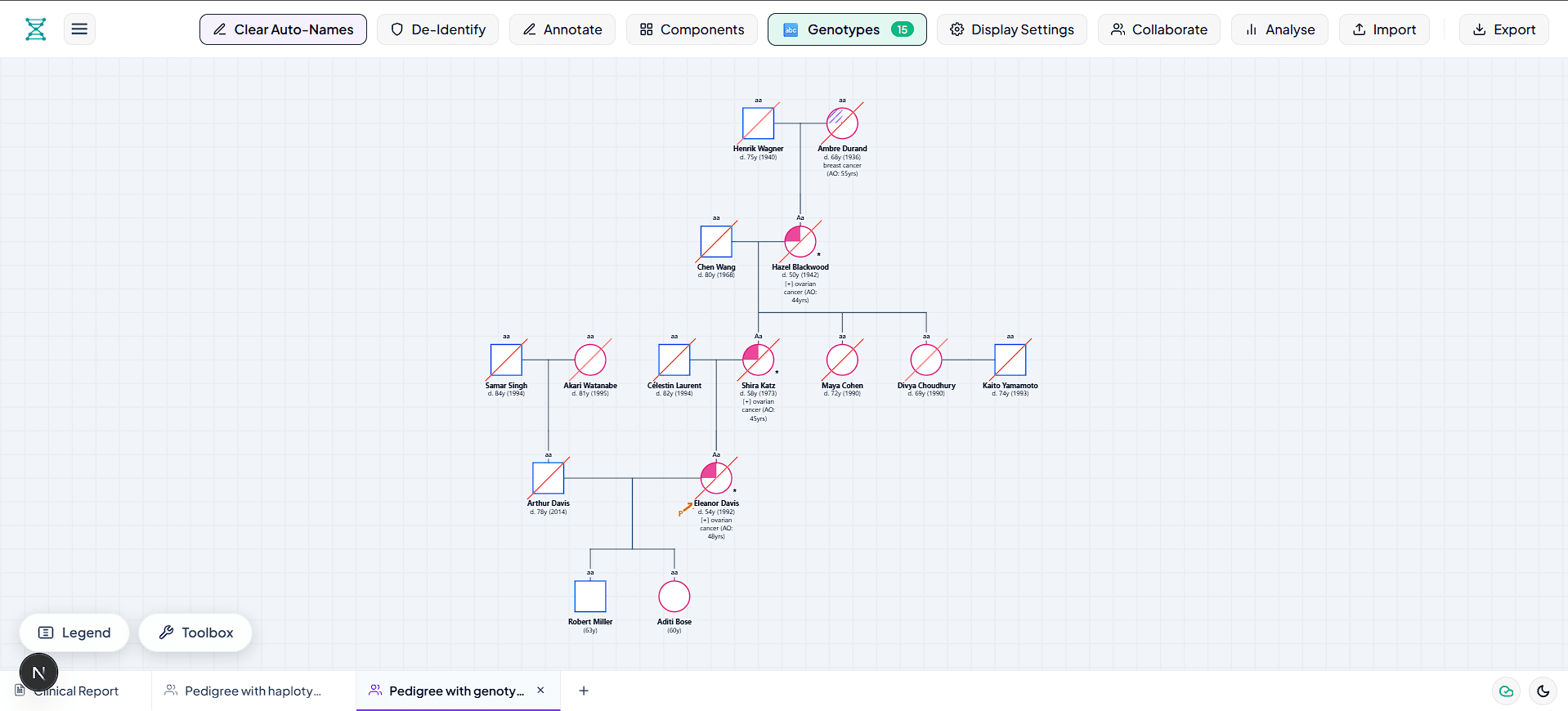
Key Takeaways
- Phenotype vs Genotype: A standard pedigree shows what a person looks like clinically (phenotype), while a pedigree chart with genotypes shows their underlying genetic makeup (alleles).
- Systematic Derivation Rules: Recessive traits always reveal their genotype first (shaded shapes are always aa). Parents of recessive children must carry at least one recessive allele (Aa).
- The Phenocopy Trap: Clinical software must distinguish between clinical symptoms and DNA test results. A patient who tests negative for a familial dominant mutation is genetically wild-type (aa), even if they have sporadic clinical symptoms.
- Adult-Onset Caveat: Young asymptomatic children in dominant cancer pedigrees should not be prematurely labeled aa without genetic testing, because they carry a 50 percent prior risk of pre-symptomatic inheritance.
- Native Biogistry Support: Biogistry natively supports customizable genotype labels with an automated 1-click Mendelian calculation algorithm that runs entirely in your local browser.
What Is a Pedigree Chart with Genotypes?
A standard pedigree chart uses symbols to represent individuals in a family tree: squares for males, circles for females, and shaded shapes for people affected by a medical condition.
However, looking at symbols alone only tells you a person's phenotype (their visible health status). It does not tell you their genotype (their actual genetic code).
A pedigree chart with genotypes (also called a genotype pedigree or pedigree with alleles) adds allele letters underneath each person's symbol. For single-gene Mendelian conditions, these labels show whether a person is:
- Homozygous Dominant (AA): Carrying two copies of the dominant allele.
- Heterozygous Carrier (Aa): Carrying one dominant and one recessive allele.
- Homozygous Recessive (aa): Carrying two copies of the recessive allele.
- Hemizygous (XᴬY or XᵃY): Males with a single X chromosome for sex-linked conditions.
- Unknown Dominant (A_): A person who displays the dominant trait, but whose second allele is uncertain without testing.
| Label Position | Pedigree Item | Content Example | Clinical Function |
|---|---|---|---|
| Top Symbol | Shape & Shading | Square with quadrant fill | Displays biological sex and observed clinical condition |
| Line 1 | Identifier & Age | II-1 (Age 48) | Individual identifier within family tree |
| Line 2 | Clinical Diagnosis & Test | [+] BRCA1 or [-] Cancer |
Diagnostic status and molecular test result |
| Line 3 | Mendelian Genotype | Aa or aa or XᴬY |
Exact allele combination for recurrence risk calculation |
Core Rules for Showing Genotypes on a Pedigree Chart
Whether you are preparing a clinical case report or solving genetics problems for USMLE or MCAT exams, follow these systematic deduction rules.
Rule 1: The Recessive Starting Point
In Autosomal Recessive inheritance (such as Cystic Fibrosis or Sickle Cell Disease):
- Every shaded (affected) individual must be homozygous recessive:
aa. - Both biological parents of an affected child must give at least one recessive allele (a). If the parents are healthy, both parents are guaranteed obligate carriers:
Aa. - Every child of an affected parent (aa) automatically receives at least one a allele.
Rule 2: The Dominant Inheritance Rules
In Autosomal Dominant inheritance (such as Huntington Disease, Marfan Syndrome, or hereditary cancer syndromes):
- Every unaffected individual lacks the disease allele and must be homozygous normal (wild-type):
aa. - Most affected individuals are heterozygous:
Aa. In rare cases where two affected parents have children, homozygous dominant (AA) genotypes often cause severe neonatal lethality (such as in Achondroplasia). - If an affected parent (Aa) pairs with an unaffected partner (aa), each child has a 50 percent probability of inheriting the disease.
Rule 3: The Unknown Second Allele (A_)
If an unaffected person in an autosomal recessive pedigree has an affected sibling, both parents are carriers (Aa × Aa). The healthy sibling has a 2/3 chance of being Aa and a 1/3 chance of being AA.
Because you cannot tell their genotype from phenotype alone without molecular testing, geneticists write their genotype as A_ (read as A-blank).
Rule 4: Sex-Linked (X-Linked) Notation
Because human males have one X chromosome and one Y chromosome (XY), they cannot be carriers for X-linked conditions:
- Normal Male:
XᴬY - Affected Male:
XᵃY(hemizygous) - Normal Female:
XᴬXᴬ - Carrier Female:
XᴬXᵃ - Affected Female:
XᵃXᵃ
Visualizing Genotypes: Two Detailed Clinical Examples
Example 1: Autosomal Dominant Pedigree with a Tested Negative Phenocopy
Below is an authentic clinical example of an Autosomal Dominant family tree.
Pay close attention to Generation I. The grandmother I-2 carries the familial pathogenic mutation (Aa) but remains clinically asymptomatic due to incomplete penetrance or sex-limited expression. The grandfather I-1 was diagnosed with late-onset prostate cancer, but his genetic test came back negative [-] for the family's mutation. Because he does not carry the familial mutation, his genotype is wild-type (aa). This is known as a phenocopy.
In Generation II, son II-3 inherited the familial mutation from his mother (Aa) and developed hereditary prostate cancer ([+]). Daughter II-2 inherited the normal maternal allele (aa) and is unaffected; she marries an unaffected partner II-1 (aa), passing normal alleles to grandson III-1 (aa).
Figure 1: Clinical Autosomal Dominant pedigree chart with genotypes. Note I-1: despite having clinical cancer, he tested negative [-] for the familial mutation, making him a sporadic phenocopy with genotype aa. Mother I-2 is the asymptomatic mutation carrier (Aa), passing the variant to affected son II-3 (Aa).
Example 2: Consanguinity and Autosomal Recessive Carrier Risk (A_)
Consanguineous matings (matings between blood relatives, such as first cousins) are represented on pedigree charts using a double horizontal marriage line.
When closely related individuals have children, rare recessive alleles carried silently in the family line have a much higher chance of meeting in homozygous form (aa).
In the pedigree below, both first cousins in Generation III are healthy carriers (Aa). They have a child in Generation IV with cystic fibrosis (aa). Notice the healthy sibling IV-2: because this sibling is unaffected, their genotype is written as A_ because they have a 2/3 prior probability of being a carrier.
Figure 2: Consanguineous Autosomal Recessive pedigree chart with standard sequential numbering (II-1 through II-4). Notice the double horizontal line between first cousins III-1 and III-2. Both cousins carry the familial mutation (Aa), producing an affected child IV-1 (aa). Healthy sibling IV-2 has genotype A_ with a 2/3 probability of being a carrier.
Clinical Molecular Nomenclature: Bridging Mendelian Letters (AA, Aa, aa) with HGVS Reports
In classroom genetics and board exams (USMLE, ABGC, MCAT), family trees are typically taught using single-letter Mendelian shorthand: AA, Aa, and aa.
However, in accredited molecular diagnostic laboratories and hospital genetics clinics, genetic test reports use HGVS (Human Genome Variation Society) sequence nomenclature. A clinical pedigree chart must often bridge both worlds: displaying simplified allele codes for family counseling while retaining the exact molecular variant for medical records.
Zygosity and Mutation Classifications
| Clinical State | Mendelian Shorthand | Molecular Classification | Clinical Lab HGVS Example | Medical Significance |
|---|---|---|---|---|
| Homozygous Wild-Type | AA (or aa in AD) |
Non-Carrier (Normal) | c.[=];[=] |
Normal diploid sequence; negative for targeted familial variant. |
| Heterozygous Pathogenic | Aa |
Heterozygote | c.5266dupC (p.Gln1756Profs*74) het |
Carries 1 mutant and 1 wild-type allele. Dominant disease or recessive carrier. |
| Homozygous Pathogenic | aa |
Homozygote | c.1521_1523delCTT (p.Phe508del) hom |
Both homologous chromosomes carry identical pathogenic mutations. |
| Compound Heterozygote | a1/a2 |
Biallelic Distinct Variants | c.[1521_1523delCTT];[3846G>A] |
Carries two distinct pathogenic variants in the same gene in trans. Recessive disease. |
| Hemizygous | XᵃY |
Hemizygote | c.40dupA (p.Tyr14*) hem |
Single X-linked mutant copy in males; lacks a homologous wild-type counterpart. |
| Variant of Uncertain Significance | A_ / VUS |
Ambiguous Variant | c.3113A>G (p.Glu1038Gly) |
Functional impact unclear; clinical management should NOT be based on VUS alone. |
The Compound Heterozygosity Challenge in Autosomal Recessive Pedigrees
In textbooks, autosomal recessive diseases are almost always depicted as homozygous aa. In clinical practice, however, many patients with recessive diseases (such as Cystic Fibrosis, Stargardt disease ABCA4, or Phenylketonuria PAH) are compound heterozygotes.
A compound heterozygote inherits one mutation from the mother (e.g., Variant 1) and a completely different mutation in the same gene from the father (e.g., Variant 2). Because both copies of the gene are disrupted, the patient manifests the full disease phenotype:
On a clinical pedigree chart, labeling a compound heterozygous child as simply aa hides the distinct molecular etiologies. Biogistry allows genetic counselors to display exact dual-variant strings beneath the symbol (such as ΔF508 / G542X or a1/a2), ensuring clinical clarity across multi-generation workups.
Bayesian Risk Calculation on Genotype Pedigrees
Pedigree charts with genotypes are not static records; they are primary mathematical tools used by genetic counselors to calculate recurrence risks and carrier probabilities.
When family test data is incomplete or disease onset is delayed by age, clinicians use Bayes' Theorem to combine standard Mendelian prior probabilities with clinical conditional information (such as age of the consultand or number of unaffected children):
Clinical Case 1: The Healthy Sibling of an Autosomal Recessive Proband
When parents have a child with Cystic Fibrosis (aa), both parents are obligate carriers (Aa × Aa). Under standard Mendelian segregation:
- 1/4 (25%) chance of
AA(unaffected non-carrier) - 1/2 (50%) chance of
Aa(unaffected carrier) - 1/4 (25%) chance of
aa(affected)
Once a sibling is born and clinically examined to be completely healthy, their probability of being aa drops to zero. Their genotype must be assigned as A_. Bayes' theorem formally calculates their carrier risk:
| Genetic Hypothesis | Prior Probability | Conditional Probability (Healthy Phenotype) | Joint Probability | Posterior Probability (Carrier Risk) |
|---|---|---|---|---|
Sibling is Non-Carrier (AA) |
1/4 (0.25) | 1.0 (Must be healthy) | 0.25 | 0.25 / 0.75 = 1/3 (33.3%) |
Sibling is Carrier (Aa) |
1/2 (0.50) | 1.0 (Must be healthy) | 0.50 | 0.50 / 0.75 = 2/3 (66.7%) |
Sibling is Affected (aa) |
1/4 (0.25) | 0.0 (Excluded by clinical observation) | 0.00 | 0% (Excluded) |
| Total / Normalizing Constant | 1.00 | - | 0.75 | 1.00 (100%) |
Clinical Case 2: Age-Dependent Penetrance in Adult-Onset Dominant Diseases
In adult-onset autosomal dominant disorders (such as Huntington disease, Early-Onset Alzheimer disease PSEN1, or hereditary cancer syndromes), an unaffected consultand's risk of carrying the mutation decreases the longer they remain symptom-free.
Suppose a 50-year-old woman (III-2) has an affected father with Huntington disease (Aa). At birth, her prior risk was 50% (1/2). The cumulative penetrance of Huntington disease by age 50 is known to be approximately 70% (0.70 of gene carriers show symptoms by age 50; 0.30 remain asymptomatic).
Because she has reached age 50 without symptoms, what is her revised probability of carrying the dominant allele (Aa)?
| Clinical Hypothesis | Prior Probability | Conditional Probability (Asymptomatic at Age 50) | Joint Probability | Posterior Probability |
|---|---|---|---|---|
Consultand carries mutation (Aa) |
1/2 (0.50) | 1 - 0.70 = 0.30 | 0.50 × 0.30 = 0.15 | 0.15 / 0.65 = 23.1% |
Consultand is wild-type (aa) |
1/2 (0.50) | 1.00 (Will never show symptoms) | 0.50 × 1.00 = 0.50 | 0.50 / 0.65 = 76.9% |
| Total / Normalizing Constant | 1.00 | - | 0.65 | 1.00 (100%) |
By remaining asymptomatic until age 50, her risk of carrying the mutation has dropped from 50% down to 23.1%. Marking her genotype as aa would be a dangerous false negative, whereas marking her as Aa would cause unwarranted anxiety. A genotype pedigree chart accurately represents her status as A_ (23% risk) or notes her age-conditioned probability until molecular genetic testing is performed.
Incomplete Penetrance, Variable Expressivity, and Non-Penetrant Obligate Carriers
When analyzing a pedigree chart with genotypes, clinicians must distinguish between two essential genetics concepts:
- Penetrance: The all-or-nothing statistical probability that a person who carries a pathogenic genotype will express any clinical symptoms of the disorder. If an allele has 80% penetrance, 20% of individuals with the disease genotype will remain completely healthy throughout their lives.
- Variable Expressivity: The range or severity of clinical signs displayed by individuals who have the exact same disease genotype. For example, in Neurofibromatosis Type 1 (NF1), one sibling with genotype
Aamay only have mild café-au-lait macules, while their sibling with the identicalAagenotype develops severe plexiform neurofibromas and scoliosis.
The Non-Penetrant Obligate Carrier Trap
A non-penetrant obligate carrier is an individual in an autosomal dominant family tree who must carry the disease variant (Aa) based on strict pedigree logic, yet exhibits zero clinical symptoms:
- An unaffected mother whose father and biological children both have an autosomal dominant condition (such as Retinoblastoma or Hereditary Paraganglioma).
- Because the condition is transmitted through her, she is an obligate carrier (
Aa).
If a genetic counselor evaluates only the visual shapes on a pedigree, an unshaded circle would suggest a healthy wild-type person (aa). This leads to catastrophic counseling errors, such as reassuring her children that their recurrence risk is negligible.
On a standard NSGC pedigree chart with genotypes, the non-penetrant obligate carrier is depicted with an unshaded circle, a center vertical dividing line or dot, and an explicit Aa (Obligate Carrier) label directly underneath.
ACMG & NSGC Standard Placement Rules for Genotypes (Bennett et al. 2008)
To avoid ambiguity in clinical records, medical journals, and courtrooms, the National Society of Genetic Counselors (NSGC) and the American College of Medical Genetics and Genomics (ACMG) established standard guidelines for annotating family trees (Bennett et al., Journal of Genetic Counseling, 2008).
When building a pedigree chart with genotypes, information must be placed directly beneath each symbol in four standardized, non-overlapping horizontal tiers:
Protocol for Recording Genetic Test Results
- Test Status Prefix: Always prepend molecular test results with brackets:
[+]indicates positive for the targeted familial mutation.[-]indicates negative for the targeted familial mutation (wild-type non-carrier).[VUS]indicates a Variant of Uncertain Significance.[Uninformative]indicates a test was performed but could not confirm or exclude pathogenicity.
- Deceased Individuals: If an individual is deceased, a single diagonal slash is drawn from the upper right corner to the lower left corner through the symbol. Their age at death and cause of death are recorded on Line 1 and Line 2 (
d. 54y, Breast Ca). - Proband Identification: The consultand or proband (the individual who brought the family to clinical attention) is identified with a solid black arrow pointing to the lower left corner of their symbol, labeled with the letter
P.
3 Clinical Traps That Most Software Gets Wrong
Most drawing tools are basic graphic editors that do not understand medical genetics. Here are three common clinical traps and how Biogistry solves them:
1. The Negative Test Result Trap (Phenocopies)
In oncology genetics, sporadic cancers are common. A grandfather might have prostate cancer due to age or environmental factors, while his children carry a hereditary BRCA2 mutation.
If the grandfather tests negative ([-]) on a genetic panel, his disease is a phenocopy. Genetically, he does not carry the family mutation. Basic software tools mistakenly assign him Aa because he is marked affected. Biogistry automatically detects the negative test result and assigns the true wild-type genotype (aa).
2. The Dominant vs Recessive Notation Clash
In an Autosomal Recessive chart, normal people are labeled AA and affected people are aa.
In an Autosomal Dominant chart, the opposite is true: normal people are aa (non-carriers) and affected people are Aa.
Many software tools use a hardcoded default that marks all healthy people as AA. This creates glaring errors where an Autosomal Dominant family tree displays affected people as Aa and healthy spouses as AA. Biogistry detects the disease inheritance pattern across the pedigree and maintains consistent notation for every relative.
3. Pre-Symptomatic Adult-Onset Risk
In conditions like Huntington disease or Lynch syndrome, symptoms do not appear until age 40 to 60.
A 20-year-old unaffected child of an Aa parent has a 50 percent prior risk of carrying the mutation. Prematurely labeling them aa based solely on their healthy appearance is clinically misleading. Biogistry lets clinicians leave genotypes blank, use the A_ chip, or tag test status until a genetic test report is finalized.
Why Biogistry Is the Superior Software for Genotype Pedigrees
Traditional clinical pedigree tools are either rigid enterprise databases or abandoned desktop programs. Biogistry modernizes the entire workflow:
| Feature / Capability | Legacy Desktop (Progeny, Cyrillic) | Diagram Software (GenoPro, SmartDraw) | Manual Drawing (Pen & Paper) | Biogistry |
|---|---|---|---|---|
| Mendelian Auto-Derive | Rule-based scripts with frequent notation errors | None (Must type text manually) | Manual calculation | 1-Click automatic derivation with clinical test overrides. |
| Negative Test Awareness | Manual workaround required | None | High risk of human error | Automatically assigns wild-type to test-negative phenocopies. |
| Quick Allele Chips | Slow dropdown menus | None | None | Instant chips (AA, Aa, aa, A_, XᴬY, XᵃY) with 1 click. |
| Privacy & Security | Device-locked desktop or cloud database | Local file or unsecured cloud | Physical paper vulnerabilities | 100% Local-First. Zero patient data sent to remote servers. |
| Mode Switching | Rebuild chart from scratch | None | Redraw entirely | Toggle between Standard, Genotypes, and Haplotypes in 1 click. |
| Export Formats | Proprietary files or low-res images | Basic image exports | Physical paper only | Lossless Vector SVG, 4K PNG, PDF, and FHIR export. |
Step-by-Step Guide: How to Create a Pedigree Chart with Genotypes in Biogistry
Creating an accurate pedigree chart with genotypes in Biogistry takes less than two minutes:
Step 1: Open Biogistry and Choose Genotypes Mode
- Launch the Biogistry Pedigree Builder.
- When creating your new file or sheet, select ** Genotypes Mode**.
- If you already have an existing family tree, simply click Display Settings and select Genotypes to reveal the genotype label layer under each symbol.
Step 2: Use 1-Click Auto-Derivation
- Click the ** Genotypes** button in the top navigation bar.
- Select your gene symbol (such as A, B, or gene name) and confirm the inheritance mode (Autosomal Dominant, Autosomal Recessive, or X-Linked).
- Click ** 1-Click Auto-Derive All**. Biogistry instantly analyzes every person, their conditions, carrier statuses, and genetic test results, filling in genotypes across the entire family.
Step 3: Fine-Tune Individual Genotypes
- Click on any person on your canvas to open the Node Inspector.
- In the Basic tab, scroll to the ** Genotype** field.
- Click any of the quick allele chips (
AA,Aa,aa,A_,XᴬY,XᵃY) or type a custom notation. - Click ** Auto-derive** on any single node to re-calculate their specific genotype at any time.
Step 4: Export Vector Figures and Clinical Reports
- Click Export in the top navigation bar.
- Export as Vector SVG for journal articles, 4K PNG for slide decks, or Clinical PDF for patient charts.
Privacy First: Complete Data Security for Clinical Genetics
Under HIPAA, GDPR, and international genetic privacy laws, uploading identifiable genetic alleles and family health histories to third-party cloud servers creates enormous compliance liabilities.
Biogistry operates on a zero-trust, local-first architecture:
- Your Data Stays on Your Machine: The pedigree engine, Mendelian calculations, and rendering algorithms execute 100 percent inside your browser's local memory.
- No Telemetry or Server Storage: Biogistry never sends patient names, family relationships, or genotype results to external servers.
- Air-Gap Friendly: Because Biogistry runs entirely in the browser, hospital genetics departments can use it in restricted clinical networks without exposing internal data to the internet.
Frequently Asked Questions (FAQ)
What is the difference between a carrier and an affected individual on a genotype pedigree?
In autosomal recessive traits, a carrier has genotype Aa. They carry one disease allele but remain healthy. An affected individual has genotype aa and shows clinical symptoms. On the pedigree chart, carriers are often marked with a center dot or half-filled symbol, while affected individuals have fully shaded symbols.
Can I display both genotypes and condition names beneath each person?
Yes. Biogistry lets you customize label lines beneath each person. You can show names, ages, diagnoses, genetic test results ([+] or [-]), and genotypes simultaneously without overlapping.
What happens if an individual's genotype is unknown?
If a person's genotype cannot be deduced from family relationships, you can leave the genotype field blank or use the A_ chip. This signals that they display the dominant trait but their second allele remains untested.
Does Biogistry support X-linked and mitochondrial genotype annotations?
Yes. Biogistry supports sex-linked alleles (XᴬY, XᵃY, XᴬXᴬ, XᴬXᵃ, XᵃXᵃ), Y-linked alleles, and mitochondrial markers (mt, mt*).
Create Your Genotype Pedigree Chart in Biogistry
Ditch the pen and paper and stop fighting with rigid drawing programs. Biogistry gives you a professional, intelligent pedigree maker with genotypes that is fast, accurate, and completely private.